Dermatologic ultrasound in hyaluronic acid removal has revolutionized the field of aesthetic medicine, providing unparalleled safety and precision. As the popularity of dermal fillers continues to soar worldwide, the need for effective, controlled reversal procedures has grown equally. Whether dealing with overfilling, asymmetry, delayed-onset nodules, or vascular compromise, the integration of advanced imaging technology ensures that corrective procedures are no longer performed blindly.
The Evolution of Filler Reversal: Why Ultrasound Changes Everything
For years, injectors relied solely on anatomical knowledge and palpation to dissolve unwanted dermal fillers. However, because hyaluronidase (the enzyme used to break down the filler) acts rapidly, precise placement is absolutely vital. Utilizing dermatologic ultrasound in hyaluronic acid removal allows practitioners to see exactly where the product is located within the soft tissue layers.
Without real-time imaging, treating complications can be a guessing game. Fillers can migrate away from the initial injection site, making physical exams misleading. By adopting dermatologic ultrasound in hyaluronic acid removal, specialists can visualize the exact depth, volume, and boundaries of the hyaluronic acid deposit, minimizing damage to surrounding healthy tissues.
### How Dermatologic Ultrasound in Hyaluronic Acid Removal Enhances Safety
Safety is the cornerstone of any aesthetic practice. When a patient presents with an adverse event or is simply unhappy with their cosmetic outcome, dermatologic ultrasound in hyaluronic acid removal provides a definitive diagnostic map. High-frequency ultrasound transducers produce high-resolution images that differentiate between skin layers, muscles, blood vessels, and foreign materials.
+------------------------+---------------------------------------+
| Traditional Removal    | Ultrasound-Guided Removal             |
+------------------------+---------------------------------------+
| Blind injections       | Real-time visual guidance             |
| Higher enzyme doses    | Targeted, lower enzyme doses          |
| Increased tissue risk  | Maximum preservation of healthy skin  |
+------------------------+---------------------------------------+
Using dermatologic ultrasound in hyaluronic acid removal drastically reduces the risk of accidental intravascular injections of hyaluronidase, which could potentially worsen a vascular crisis. It gives both the patient and the physician immense peace of mind.
Step-by-Step: The Ultrasound-Guided Dissolving Process
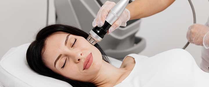
The actual procedure involving dermatologic ultrasound in hyaluronic acid removal is highly efficient. First, the clinician applies a sterile gel to the target area and sweeps the ultrasound probe across the skin. The hyaluronic acid appears as a distinct echoic structure—often anechoic (dark) or hypoechoic—trapped within the tissue layers.
Once identified, the needle is introduced into the skin under direct, continuous ultrasound visualization. The practitioner can watch the needle tip enter the exact center of the filler deposit. This level of accuracy means that dermatologic ultrasound in hyaluronic acid removal requires significantly less hyaluronidase, reducing post-treatment swelling and allergic reactions.
### Addressing Complications with Dermatologic Ultrasound in Hyaluronic Acid Removal
When severe complications like vascular occlusion occur, every second counts. The immediate application of dermatologic ultrasound in hyaluronic acid removal protocols allows doctors to locate the precise area of compression or blockage. This rapid intervention can prevent skin necrosis and long-term scarring.
Even in non-emergency cases, such as chronic swelling or late-onset granulomas, dermatologic ultrasound in hyaluronic acid removal helps distinguish between an inflammatory response to the filler and a bacterial infection. This distinction alters the treatment plan entirely, shifting from antibiotics to targeted enzyme dissolution.
Medical Tourism and Advanced Aesthetic Care
Patients looking for top-tier aesthetic corrections often travel internationally to find clinics equipped with this state-of-the-art technology. If you are considering combining advanced medical procedures with world-class hospitality, exploring options for Plastic Surgery in Brazil is an excellent choice, as the country boasts some of the most strictly trained ultrasound-guided injectors in the world.
Clinics dedicated to excellence ensure that every corrective procedure is backed by rigorous imaging protocols. If you want to see the real-world impact of these advanced cosmetic interventions, you can Check out the results of some patients. to understand how precision mapping restores natural facial harmony.
Choosing the Right Technology and Clinic
Not all ultrasound machines are suitable for facial aesthetics. High-frequency probes (usually 15 to 22 MHz or higher) are mandatory to capture the superficial layers of the face clearly. When clinics invest in this advanced machinery, it reflects a deep commitment to patient care and successful outcomes.
For comprehensive information regarding cutting-edge facial treatments, dermatologic care, and advanced filler management, you can visit the official website of Belvivere, a leading institution specializing in premium aesthetic medicine and patient safety.
The Future of Aesthetic Dermatology
The integration of imaging tech is no longer just an optional luxury; it is quickly becoming the global standard of care. Relying on dermatologic ultrasound in hyaluronic acid removal ensures that the field of aesthetics moves away from subjective assessments and moves toward objective, data-driven medicine.
As fillers continue to evolve with different cross-linking technologies, their degradation behaviors change too. Having a reliable tool like dermatologic ultrasound in hyaluronic acid removal ensures that no matter how complex the filler chemistry is, the removal process remains safe, predictable, and highly successful for every patient involved.
In conclusion, prioritizing dermatologic ultrasound in hyaluronic acid removal protects the patient’s anatomy, optimizes the enzyme’s efficiency, and guarantees a superior standard of medical care that eliminates guesswork entirely.